Capsule Endoscopy involves swallowing a video capsule, the size of a large vitamin capsule, to examine primarily the small bowel, middle part of your gastrointestinal tract, which cannot be easily examined by traditional endoscopy methods (upper endoscopy or by ileo-colonoscopy). The procedure is commonly undertaken if there is suspicion of non-stricturing disease of the small bowel. The capsule endoscopy is a visual examination and is purely diagnostic, no biopsies or therapy can be performed with this.
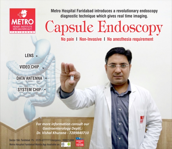
This capsule, with its own camera and light source, takes pictures of small intestine as it passes through and transmits it to small recording device which is wear as belt on body. These images are seen real time or later on.
Why is Capsule Endoscopy Done?
Capsule endoscopy helps to evaluate the small intestine. The capsule endoscopy is indicated to search for source of small intestinal bleeding and to detect polyps, inflammatory bowel disease (Crohn’s disease), tumors and ulcers of the small intestine.
What preparation is required for Capsule endoscopy?
Usually 12 hours fasting is recommended and sometimes bowel preparation/cleansing may be done prior to the examination.
How Capsule Endoscopy is done?
The pill-sized capsule endoscope is swallowed with a small amount of water. Sensors (like ECG leads) are placed on the patient's abdomen. The sensors will be attached to a Data Recorder that will be worn in a belt around the patient's waist. Once swallowed the camera moves naturally through the digestive tract. Approximately eight hours after ingesting the camera, patients return to the Endoscopy Unit where the recording device and the sensors are removed, the images are downloaded to a computer and evaluated. This is disposable/single time use capsule and it passes of its own in stool. The patient are advised to check the passage of the capsule in their stool regularly and to report it to the Physician. Patient is instructed not to undergo MRI till capsule is inside their body.
What are its benefits?
The capsule is easily swallowed and non-invasive. It is painless, real time and sedation free. Patient can relax in comfort and walk about, without a hospital stay. Exposure to potentially harmful radiation does not occur
Capsule Endoscopy is usually used when other methods such as: Upper GI endoscopy (gastroduodenoscopy), Colonoscopy and/or Enteroscopy have failed to provide a diagnosis.
Alternative to capsule endoscopy is Balloon endoscopy (single/double balloon) which involves passing a long endoscope into the small bowel which is more invasive, requires sedation and carries a greater risk.
What Happens After Capsule Endoscopy?
After capsule ingestion patient is allowed drink clear liquid after 2 hours and given soft diet after 4hours, unless doctor instructs otherwise. Patient can perform routine daily activity but should avoid vigorous physical activity during the study.
What are the Possible Complications of Capsule Endoscopy?
Although complications can occur, they are rare. The main complications following a capsule endoscopy are mentioned below. In approximately 1-2 out of 100 procedures, the capsule can become lodged above a stricture (narrowing) of the digestive tract. Patients who have Crohn’s disease or have had abdominal surgery in the past are at increased risk for this complication. An abdominal x-ray may be ordered if the physician is not able to determine that the capsule passed into the large intestine during the course of the study.
Dr Vishal khurana
MBBS, MD, DM (Gastroenterology), MNAMS
Senior consultant- Gastroenterology
Metro hospital, Faridabad


 Appointment
 Find A Doctor
 Lab Report